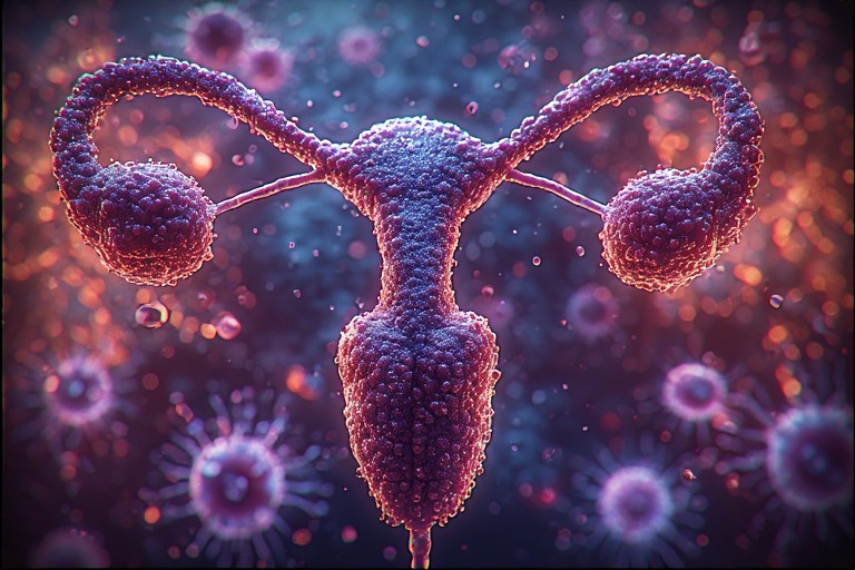
How Endocrine Disruptors, Hidden Infections, and Immune Dysregulation Silently Destroy Reproductive Capacity
The 59% decline in sperm counts across Western nations over four decades is not idiopathic—it is the measurable consequence of ubiquitous endocrine-disrupting chemical exposure, with bisphenol A detected in 93% of Americans and phthalates in 98% of the population. These compounds operate at molecular concentrations, binding hormone receptors, suppressing testosterone synthesis, disrupting folliculogenesis, and impairing implantation through mechanisms that standard fertility evaluations never assess. Simultaneously, hidden infections (Chlamydia, Ureaplasma, Mycoplasma, chronic viral burden) produce tubal scarring, sperm DNA damage, and chronic inflammation, while immunological factors including antiphospholipid antibodies and elevated natural killer cells attack implanting embryos. This article examines the pathophysiological mechanisms through which these hidden causes sabotage reproductive function at every level—from hypothalamic signaling to implantation.
Key Clinical Takeaways
- BPA is found in 93% of Americans and is linked to 20% lower IVF implantation rates — bisphenol A binds estrogen receptors as a functional xenoestrogen, disrupting follicular development, impairing endometrial receptivity, and reducing sperm concentration at concentrations commonly detected in human serum
- Phthalates suppress testosterone and reduce sperm count in 98% of the population — these plasticizers inhibit Leydig cell steroidogenesis, reducing fetal and adult testosterone production; metabolite levels correlate directly with diminished sperm parameters and increased time to pregnancy
- PFAS (“forever chemicals”) reduce fertility rates by 16–21% — per- and polyfluoroalkyl substances persist in tissue for years, disrupt ovarian function, alter steroidogenesis, and are associated with longer time to pregnancy and reduced live birth rates
- Hidden infections cause tubal scarring and sperm DNA damage without obvious symptoms — Chlamydia trachomatis, Ureaplasma urealyticum, and Mycoplasma hominis produce subclinical pelvic inflammatory disease, tubal factor infertility, and direct sperm DNA fragmentation through oxidative stress mechanisms
- Cortisol “steals” from the progesterone pathway under chronic stress — elevated cortisol dysregulates the hypothalamic-pituitary-adrenal axis, shunting pregnenolone away from progesterone synthesis and toward cortisol production, creating functional luteal phase deficiency
The Endocrine Disruption Epidemic: Fertility Under Molecular Siege
The average reproductive-age woman in an industrialized nation carries measurable body burdens of more than 100 synthetic chemicals, many of which possess endocrine-disrupting properties. These compounds are not theoretical hazards—they are present in blood, urine, follicular fluid, amniotic fluid, and breast milk at concentrations sufficient to alter hormonal signaling.
The decline in reproductive parameters over the past four decades is not mysterious. It is the predictable outcome of saturating a biological system with compounds that interfere with the most hormone-sensitive process in human physiology: reproduction.

Bisphenol A: The Universal Xenoestrogen
Bisphenol A (BPA) is one of the highest-volume chemicals produced worldwide, used in polycarbonate plastics, epoxy resins, thermal paper, and food packaging. The Centers for Disease Control and Prevention’s National Health and Nutrition Examination Survey (NHANES) has consistently detected BPA in approximately 93% of the U.S. population.
BPA’s reproductive toxicity operates through multiple mechanisms:
- Estrogen receptor binding: BPA binds both ERα and ERβ with affinity sufficient to activate estrogenic signaling at concentrations well below those currently considered “safe” by regulatory agencies. In the ovary, this disrupts follicular development and oocyte maturation. In the testis, it alters Sertoli cell function and spermatogenesis.
- Anti-androgenic activity: BPA antagonizes androgen receptor signaling, reducing testosterone’s effects on spermatogenesis and male sexual development. In male fetuses, BPA exposure during critical windows of sexual differentiation is associated with shortened anogenital distance—a marker of androgen insufficiency that correlates with reduced adult semen quality.
- Epigenetic reprogramming: Prenatal BPA exposure produces heritable epigenetic modifications that alter gene expression in subsequent generations. Animal models demonstrate that grandmaternal BPA exposure reduces fertility in granddaughters—an intergenerational toxicity that compounds with each successive exposure.
- IVF implantation disruption: A prospective study of women undergoing IVF found that those with the highest urinary BPA concentrations had approximately 20% lower implantation rates compared to those with the lowest levels. The mechanism involves impaired endometrial receptivity—BPA alters the expression of integrins and other adhesion molecules essential for embryo implantation.
The “BPA-free” label provides no reassurance. Bisphenol S (BPS) and bisphenol F (BPF), the common replacements, exhibit comparable or even greater estrogenic activity in vitro. The substitution replaces one endocrine disruptor with another while maintaining the consumer’s exposure.
Phthalates: The Testosterone Suppressors
Phthalates—plasticizers used in vinyl flooring, food packaging, personal care products, and medical devices—are detected in 98% of the population. Their reproductive toxicity is predominantly anti-androgenic.

Di(2-ethylhexyl) phthalate (DEHP) and its metabolite mono-(2-ethylhexyl) phthalate (MEHP) directly inhibit Leydig cell steroidogenesis, reducing testosterone production at the cellular level. The consequence is dose-dependent: higher phthalate metabolite concentrations in urine correlate with lower sperm concentration, reduced motility, and altered morphology.
In women, phthalate exposure is linked to polycystic ovary syndrome (PCOS), endometriosis, and implantation failure. The mechanism involves disruption of ovarian steroidogenesis, alteration of follicle-stimulating hormone signaling, and interference with the progesterone receptor activation necessary for endometrial decidualization.
The critical window of vulnerability for phthalate exposure begins in utero. Male fetuses exposed to high maternal phthalate concentrations during the first trimester—the period of testicular differentiation and initial spermatogonial stem cell development—demonstrate measurable reductions in anogenital distance and subsequent reproductive parameters.
PFAS: The Forever Chemicals That Persist in Reproductive Tissue
Per- and polyfluoroalkyl substances (PFAS)—used in non-stick cookware, water-resistant fabrics, food packaging, and firefighting foam—resist degradation in both the environment and the human body. The half-life of some PFAS compounds in human serum exceeds five years.
Epidemiological data consistently associate PFAS exposure with reduced fertility:
- 16–21% lower fertility rates in women with higher serum PFAS concentrations
- Longer time to pregnancy in couples where either partner has elevated PFAS levels
- Reduced ovarian reserve as measured by lower antral follicle counts and diminished AMH levels
PFAS disrupt reproductive function through interference with ovarian steroidogenesis, altered FSH and LH signaling, impaired oocyte quality, and disruption of placental development. Their persistence means that exposure occurring years before conception may still be present at biologically active concentrations during the critical peri-conception period.
Heavy Metals: Placental Crossers With No Safe Threshold
Heavy metals represent a distinct category of reproductive toxicant because they cross the placental barrier with particular efficiency and have no established safe threshold for fetal exposure.
- Lead: Crosses the placenta freely; accumulates in fetal tissue; associated with miscarriage at any detectable level. Even blood lead concentrations previously considered “normal” are associated with increased spontaneous abortion risk and reduced fecundability.
- Mercury: Methylmercury concentrates in the fetal brain, producing neurological damage. For the mother, mercury exposure impairs ovarian function and alters hormone metabolism. The primary exposure routes—fish consumption and dental amalgams—create a clinical tension between omega-3 benefits and mercury risk that requires strategic navigation.
- Cadmium: Damages ovarian function directly, reducing follicular reserve and impairing steroidogenesis. Cadmium also disrupts zinc-dependent enzyme systems critical for DNA synthesis and repair in both oocytes and sperm.
- Arsenic: Associated with increased miscarriage risk and reduced birth weight. Arsenic exposure through contaminated groundwater affects millions of reproductive-age women globally.
Pesticides and Glyphosate: The Agricultural Assault
Women consuming produce with high pesticide residues demonstrate a 26% lower probability of pregnancy compared to those consuming low-pesticide produce, according to data from the Nurses’ Health Study II. The mechanism is multifold:
- Hormone disruption: Many pesticides function as endocrine disruptors, binding estrogen, androgen, or thyroid receptors
- Gut microbiome destruction: Glyphosate’s antimicrobial properties disrupt the intestinal microbiome, which plays a critical role in estrogen metabolism, immune regulation, and nutrient absorption
- Mitochondrial toxicity: Organophosphate pesticides impair mitochondrial function in both oocytes and sperm, reducing energy production essential for fertilization and early embryo development
Hidden Infections: The Silent Scarring and DNA Damage
Standard fertility workups typically assess for active pelvic infection through basic cervical cultures or chlamydia antibody testing. This approach misses both chronic low-grade infections and organisms that produce reproductive damage without acute symptoms.
Chlamydia trachomatis: The Tubal Destroyer
Chlamydia is the most common bacterial sexually transmitted infection, and its reproductive consequences are disproportionately severe because it is frequently asymptomatic. Approximately 70–90% of chlamydial infections in women and 50–90% in men produce no acute symptoms, meaning the infection persists untreated.
The primary fertility mechanism is tubal scarring. Chlamydia triggers a chronic inflammatory response in the fallopian tube mucosa, leading to fibrosis, ciliary destruction, and tubal occlusion. The damage is progressive—each untreated infection episode increases the severity of tubal pathology.
Chlamydia antibody testing (CAT) provides indirect evidence of prior exposure but does not distinguish between treated past infection and ongoing chronic infection. Comprehensive evaluation requires PCR testing for active infection, and in some cases, laparoscopic assessment of tubal integrity.
Ureaplasma and Mycoplasma: The Overlooked Pathogens
Ureaplasma urealyticum and Mycoplasma hominis are genital mycoplasmas that colonize the reproductive tract in significant proportions of the population. Their pathogenicity has been debated, but accumulating evidence demonstrates their role in:
- Sperm DNA damage: Ureaplasma infection increases reactive oxygen species production in semen, directly damaging sperm DNA integrity
- Poor semen quality: Reduced motility and altered morphology in colonized men
- Miscarriage: Association with early pregnancy loss through subclinical endometritis and immune activation
- Preterm birth: A well-established association that suggests these organisms alter the maternal-fetal immune interface
These organisms are not detected on standard cervical cultures or routine semen analysis. Specific PCR testing is required, and it is rarely ordered in standard fertility evaluations.
Chronic Viral Burden and Immune Activation
Chronic viral infections—including herpesviruses, cytomegalovirus, and in endemic regions, Borrelia burgdorferi (Lyme disease)—can produce persistent immune activation that disrupts reproductive function. The mechanism is indirect: chronic immune stimulation elevates inflammatory cytokines (TNF-α, IL-6, IFN-γ) that interfere with implantation, alter endometrial receptivity, and create a hostile environment for embryo development.
Lyme disease deserves specific mention because its prevalence in endemic regions and its capacity for chronic, multi-system involvement make it a plausible contributor to unexplained reproductive failure. The spirochete can persist in tissue despite antibiotic treatment, and the ongoing immune response—including cross-reactive antibodies—may interfere with reproductive immunology at the implantation site.
Immunological Factors: When the Immune System Attacks Conception
The maternal immune system must accomplish a paradox: it must tolerate the implantation of a semi-allogeneic embryo (genetically half-foreign) while maintaining defense against pathogens. When this delicate immunological balance fails, the result is recurrent implantation failure or recurrent pregnancy loss.
Antiphospholipid Antibody Syndrome
Antiphospholipid antibodies (aPL)—including anticardiolipin antibodies, lupus anticoagulant, and anti-β2 glycoprotein I—interfere with placental development through multiple mechanisms:
- Thrombosis of placental vessels: aPL promote coagulation at the maternal-fetal interface, restricting blood flow to the developing placenta
- Inhibition of trophoblast invasion: aPL directly impair the ability of the embryo’s trophoblast cells to invade the endometrium and establish vascular connections
- Complement activation: aPL trigger inflammatory complement cascades at the implantation site
Antiphospholipid antibody testing is not uniformly included in standard recurrent pregnancy loss evaluations, and when performed, it often uses incomplete panels that miss some antibody subtypes.
Elevated Natural Killer Cells
Natural killer (NK) cells are lymphocytes that participate in innate immune defense. In the context of reproduction, elevated peripheral NK cell activity or abnormal uterine NK cell populations may attack the implanting embryo, recognizing trophoblast cells as abnormal and initiating cytotoxic responses.
The relationship between NK cells and reproductive failure is complex and remains debated in the literature, but clinical observations suggest that a subset of women with recurrent implantation failure or recurrent miscarriage demonstrate NK cell profiles that may benefit from immunomodulatory intervention.
Antisperm Antibodies
Antisperm antibodies—produced by either partner—can cause sperm agglutination, impair capacitation, block sperm-egg binding, or activate complement-mediated sperm damage. These antibodies are rarely tested in standard evaluations despite their capacity to interfere with fertilization at multiple stages.
The Cortisol-Progesterone Steal: Stress as a Reproductive Saboteur
The hypothalamic-pituitary-adrenal (HPA) axis and the hypothalamic-pituitary-gonadal (HPG) axis are not independent systems—they share a common precursor and a competitive metabolic pathway.
Pregnenolone is the precursor for both cortisol and progesterone. Under conditions of chronic stress—whether physiological (infection, inflammation, sleep deprivation, toxin burden) or psychological—the HPA axis upregulates cortisol production. The biosynthetic pathway is shunted: pregnenolone is preferentially converted to cortisol rather than progesterone.
This “pregnenolone steal” (more precisely, a competitive diversion of substrate) produces a functional progesterone deficiency even when luteal function appears adequate on surface assessment. The consequences cascade:
- Luteal phase deficiency: Inadequate progesterone to maintain the endometrium in a receptive state
- Impaired implantation: Progesterone is essential for endometrial decidualization and immune modulation at the implantation site
- Increased miscarriage risk: Progesterone withdrawal triggers menstrual shedding; insufficient luteal support leads to early pregnancy loss
- Sleep disruption compounds the problem: Testosterone production peaks during sleep, and sleep apnea devastates hormonal health. Melatonin—suppressed by artificial light exposure—directly modulates reproductive hormone secretion and possesses antioxidant properties essential for oocyte protection
Chronic stress does not merely “affect” fertility—it mechanically disrupts the hormonal pathways that enable conception and pregnancy maintenance. This is not a psychological observation; it is a biochemical mechanism.
The Cumulative Burden: Additive and Synergistic Effects
The most critical aspect of environmental reproductive toxicity is not any single exposure—it is the cumulative, interactive burden. Endocrine disruptors do not operate in isolation. BPA, phthalates, PFAS, heavy metals, pesticides, and chronic infections create additive and synergistic effects that exceed the impact of any individual toxin.
A woman with moderate BPA exposure, borderline phthalate levels, a subclinical chlamydial infection, and a TSH of 3.2 mIU/L does not have four minor problems. She has a compounded reproductive impairment that may exceed the sum of its parts—each factor amplifying the others through shared pathways of inflammation, oxidative stress, and endocrine disruption.
Standard fertility evaluation assesses none of these factors. The patient is declared “unexplained” and directed toward IVF, which bypasses rather than resolves the underlying sabotage.
References
- Levine H, et al. Temporal Trends in Sperm Count: A Systematic Review and Meta-Regression Analysis. Hum Reprod Update. 2017;23(6):646-659. doi:10.1093/humupd/dmx022
- Souter I, et al. Bisphenol A and IVF Outcomes: A Prospective Cohort Study. Hum Reprod. 2018;33(10):1886-1894. doi:10.1093/humrep/dey283
- Buck Louis GM, et al. Perfluorochemicals and Human Fecundity: The LIFE Study. Environ Health Perspect. 2018;126(6):067012. doi:10.1289/EHP2518
- Haahr T, et al. Reproductive Outcome of Women With Ureaplasma urealyticum Infection: A Systematic Review. Fertil Steril. 2021;116(4):1092-1100. doi:10.1016/j.fertnstert.2021.06.014
- Cappelletti M, et al. Natural Killer Cells in Reproductive Immunology: A Systematic Review. J Reprod Immunol. 2023;152:103672. doi:10.1016/j.jri.2023.103672
Medical Disclaimer
This article is intended for educational and informational purposes only. It does not constitute medical advice, diagnosis, or treatment recommendations. Always consult with a qualified healthcare provider before initiating, modifying, or discontinuing any medical intervention. The information presented reflects current evidence and clinical observations but should not replace individualized medical care.
For structured protocols and implementation guidance, visit Human Optimization Lab.